
പ്രമാണം:PLoSBiol4.e126.Fig6fNeuron.jpg

ഈ പ്രിവ്യൂവിന്റെ വലിപ്പം: 687 × 600 പിക്സലുകൾ. മറ്റ് റെസലൂഷനുകൾ: 275 × 240 പിക്സലുകൾ | 550 × 480 പിക്സലുകൾ | 915 × 799 പിക്സലുകൾ.
പൂർണ്ണ വലിപ്പം (915 × 799 പിക്സൽ, പ്രമാണത്തിന്റെ വലിപ്പം: 787 കെ.ബി., മൈം തരം: image/jpeg)
| ഈ പ്രമാണം വിക്കിമീഡിയ കോമൺസിൽ നിന്നുള്ളതാണ്. പ്രമാണത്തെക്കുറിച്ചുള്ള വിവരണം താഴെ കൊടുത്തിരിക്കുന്നു.
|
| വിവരണം |
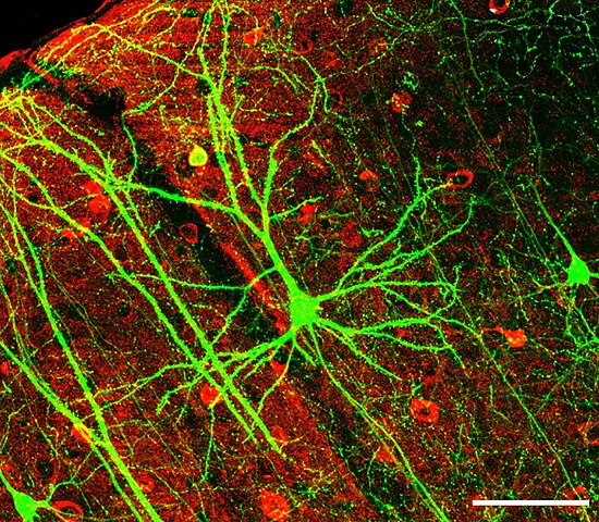
English: After the original figure legend: Coronal section containing the chronically imaged pyramidal neuron “dow” (visualized by green GFP) does not stain for GABA (visualized by antibody staining in red). Confocal image stack, overlay of GFP and GABA channels. Scale bar: 100 μm
Deutsch: Mikroskopische Aufnahme eines Pyramiden-Neurons der Maus (Zerebraler Cortex, das Grün fluoreszierendes Protein exprimiert. Die rote Antikörper-Färbung zeigt GABA-produzierende Interneuronen. Maßstabsbalken: 100 µm |
||
| തീയതി | |||
| സ്രോതസ്സ് | Dynamic Remodeling of Dendritic Arbors in GABAergic Interneurons of Adult Visual Cortex. Lee WCA, Huang H, Feng G, Sanes JR, Brown EN, et al. PLoS Biology Vol. 4, No. 2, e29. doi:10.1371/journal.pbio.0040029, Figure 6f, slightly altered (plus scalebar, minus letter "f".) | ||
| സ്രഷ്ടാവ് | Wei-Chung Allen Lee, Hayden Huang, Guoping Feng, Joshua R. Sanes, Emery N. Brown, Peter T. So, Elly Nedivi | ||
| അനുമതി (ഈ പ്രമാണത്തിന്റെ പുനരുപയോഗം) |
|
||
| മറ്റു പതിപ്പുകൾ | en:Image:GFPneuron.png |
പ്രമാണ നാൾവഴി
ഏതെങ്കിലും തീയതി/സമയ കണ്ണിയിൽ ഞെക്കിയാൽ പ്രസ്തുതസമയത്ത് ഈ പ്രമാണം എങ്ങനെയായിരുന്നു എന്നു കാണാം.
| തീയതി/സമയം | ലഘുചിത്രം | അളവുകൾ | ഉപയോക്താവ് | അഭിപ്രായം | |
|---|---|---|---|---|---|
| നിലവിലുള്ളത് | 10:34, 13 ഫെബ്രുവരി 2013 | | 915 × 799 (787 കെ.ബി.) | Hic et nunc | Maßstab wieder rein |
| 07:17, 13 ഫെബ്രുവരി 2013 |  | 921 × 805 (836 കെ.ബി.) | Hic et nunc | watermark removed | |
| 21:30, 31 ജനുവരി 2008 |  | 922 × 806 (804 കെ.ബി.) | Dietzel65 | {{Information |Description={en|After the original figure legend: Coronal section containing the chronically imaged pyramidal neuron “dow” (visualized by green GFP) does not stain for GABA (visualized by antibody staining in red). Confocal image stack, |
പ്രമാണത്തിന്റെ ഉപയോഗം
താഴെ കാണുന്ന താളിൽ ഈ ചിത്രം ഉപയോഗിക്കുന്നു:
പ്രമാണത്തിന്റെ ആഗോള ഉപയോഗം
താഴെ കൊടുത്തിരിക്കുന്ന മറ്റ് വിക്കികൾ ഈ പ്രമാണം ഉപയോഗിക്കുന്നു:
- als.wikipedia.org സംരംഭത്തിലെ ഉപയോഗം
- ar.wikipedia.org സംരംഭത്തിലെ ഉപയോഗം
- as.wikipedia.org സംരംഭത്തിലെ ഉപയോഗം
- azb.wikipedia.org സംരംഭത്തിലെ ഉപയോഗം
- cs.wikipedia.org സംരംഭത്തിലെ ഉപയോഗം
- de.wikipedia.org സംരംഭത്തിലെ ഉപയോഗം
- de.wikibooks.org സംരംഭത്തിലെ ഉപയോഗം
- Natur und Technik für den Pflichtschulabschluss: Das Leben
- Natur und Technik für den Pflichtschulabschluss: Die Evolution der Zelle
- Natur und Technik für den Pflichtschulabschluss: Neuron
- Natur und Technik für den Pflichtschulabschluss: Menschliche Gewebe
- Benutzer:Yomomo/ NuT
- Natur und Technik für den Pflichtschulabschluss/ Buch
- de.wikiversity.org സംരംഭത്തിലെ ഉപയോഗം
- de.wiktionary.org സംരംഭത്തിലെ ഉപയോഗം
- en.wikipedia.org സംരംഭത്തിലെ ഉപയോഗം
- en.wikiquote.org സംരംഭത്തിലെ ഉപയോഗം
- es.wikibooks.org സംരംഭത്തിലെ ഉപയോഗം
- fa.wikipedia.org സംരംഭത്തിലെ ഉപയോഗം
- fr.wikiversity.org സംരംഭത്തിലെ ഉപയോഗം
- gd.wikipedia.org സംരംഭത്തിലെ ഉപയോഗം
- gl.wikipedia.org സംരംഭത്തിലെ ഉപയോഗം
- hi.wikipedia.org സംരംഭത്തിലെ ഉപയോഗം
- hy.wikipedia.org സംരംഭത്തിലെ ഉപയോഗം
- ka.wikipedia.org സംരംഭത്തിലെ ഉപയോഗം
- kn.wikipedia.org സംരംഭത്തിലെ ഉപയോഗം
- ko.wikipedia.org സംരംഭത്തിലെ ഉപയോഗം
- mn.wikipedia.org സംരംഭത്തിലെ ഉപയോഗം
- ms.wikipedia.org സംരംഭത്തിലെ ഉപയോഗം
- ne.wikipedia.org സംരംഭത്തിലെ ഉപയോഗം
- nn.wikipedia.org സംരംഭത്തിലെ ഉപയോഗം
- outreach.wikimedia.org സംരംഭത്തിലെ ഉപയോഗം
ഈ പ്രമാണത്തിന്റെ കൂടുതൽ ആഗോള ഉപയോഗം കാണുക.